| 일 | 월 | 화 | 수 | 목 | 금 | 토 |
|---|---|---|---|---|---|---|
| 1 | 2 | 3 | 4 | 5 | 6 | 7 |
| 8 | 9 | 10 | 11 | 12 | 13 | 14 |
| 15 | 16 | 17 | 18 | 19 | 20 | 21 |
| 22 | 23 | 24 | 25 | 26 | 27 | 28 |
| 29 | 30 |
- 방사선사나라
- receive bandwidth
- slice gap
- TR TE
- MRI image parameters
- MRA
- T2 이완
- fractional echo
- 자기공명혈관조영술
- chemical shift artifact
- aliasing artifact
- MRI gantry
- 사전포화펄스
- 동위상 탈위상
- T1WI
- no phase wrap
- saturation band
- T2WI
- ECG gating
- wrap around artifact
- tof
- T2강조영상
- FSE
- MR angiography
- MRI 영상변수
- T1강조영상
- K-space
- radiographer nara
- saturation pulse
- fast spin echo
- Today
- Total
방사선사나라 Radiographer Nara
[MRI] (영/한) Proton density weighted image - 양자밀도 강조영상 본문
(영어/영문/English)
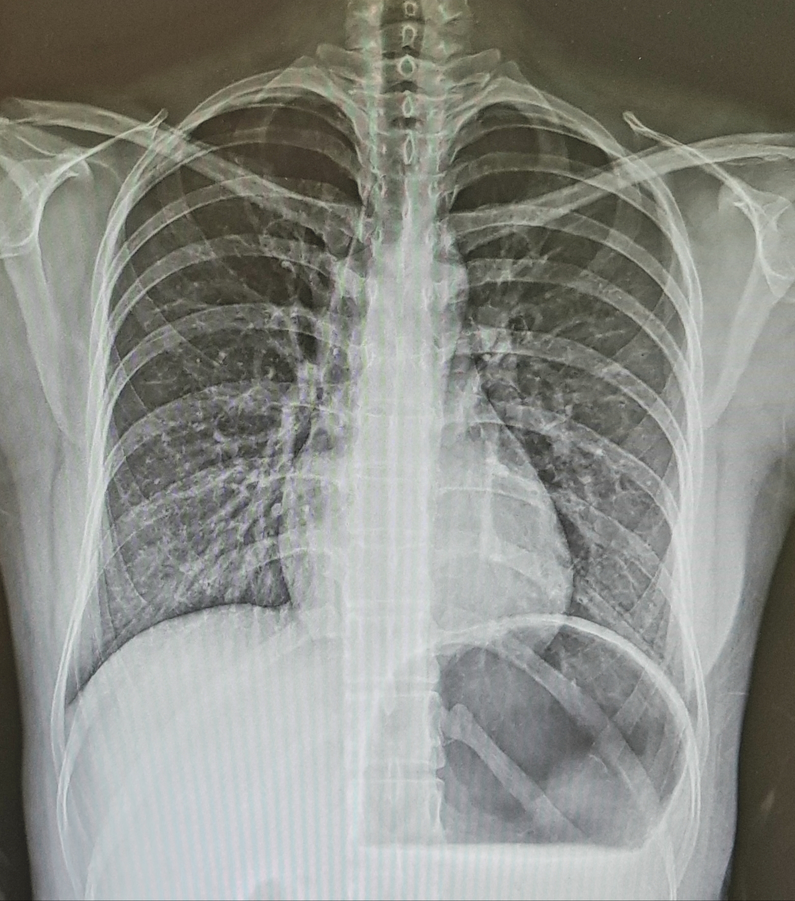
The proton density weighted image is an image that obtains a contrast proportional to the number of protons in tissue existing within a patient.
This image has the highest signal intensity and is mainly used to observe a wide range of tissues.
When the proton density weighted image minimizes the T1 and T2 components as much as possible, a true proton density weighted image is obtained. In order to exclude the T1 and T2 components, TR and TE must be selected well.
If the TR is shortened, the T1 contrast between the two tissues shows a signal with a large difference. Therefore, the T1 component should be excluded by applying a long TR. (TR is 1000ms or more)
In addition, in order to minimize the T2 component, TE should be shortened. (Clinically use about 10 ~ 30ms)
The image of the signal obtained has little difference in contrast between tissues, and is used when viewing the clinically wide area because the image is bright overall.
In conclusion, in long TR, the longitudinal magnetization is almost recovered so the T1 component can be reduced, and the short TE can reduce the T2 component.
In general, since the proton density weighted image has a long TR and many slices, a method of selecting two TEs and simultaneously obtaining a proton density weighted image and a T2 weighted image is used.

by radiographer nara
(국어/국문/Korean)
양자밀도 강조영상은 환자의 일정 범위 속에 존재하는 원자핵 즉 조직의 양자 수에 비례한 대조도를 얻는 영상으로 이 영상은 신호 강도가 가장 커서 넓은 범위의 조직들을 관찰하는데 주로 이용된다.
양자밀도 강조영상은 T1, T2 성분을 최대한 최소화했을 때 진정한 양자밀도 강조영상은 얻게 되는데 T1과 T2 성분을 배제하기 위해서는 TR과 TE를 잘 선택해야 한다.
만약 TR을 짧게 하면 두 조직의 T1 대조도 차이가 큰 신호를 나타내게 되므로 TR을 길게 인가해 T1 성분을 배제시켜야 한다. (TR은 1000ms 이상)
또한 T2 성분을 최소화하기 위해서는 TE를 어느정도 짧게 해야 한다. (임상적으로 10~30ms 정도를 이용)
이렇게 얻은 신호의 영상은 조직간의 대조도 차이는 적고 영상이 전체적으로 밝게 보여 임상적으로 넓은 영역을 볼 때 이용되고 있다.
결론적으로 긴 TR에서는 종축자기화가 거의 회복되므로 T1 성분을 감소시킬 수 있고 짧은 TE는 T2 성분을 감소시킬 수 있다.
일반적으로 양자밀도 강조영상은 TR이 길고 단면 수가 많기 때문에, 두 개의 TE를 선택해 양자밀도 강조영상과 T2 강조영상을 동시에 얻는 방법을 이용한다.
- 방사선사나라


