| 일 | 월 | 화 | 수 | 목 | 금 | 토 |
|---|---|---|---|---|---|---|
| 1 | 2 | 3 | 4 | |||
| 5 | 6 | 7 | 8 | 9 | 10 | 11 |
| 12 | 13 | 14 | 15 | 16 | 17 | 18 |
| 19 | 20 | 21 | 22 | 23 | 24 | 25 |
| 26 | 27 | 28 | 29 | 30 |
- 자기공명혈관조영술
- TR TE
- 사전포화펄스
- chemical shift artifact
- aliasing artifact
- slice gap
- MRI image parameters
- no phase wrap
- 방사선사나라
- MRI gantry
- radiographer nara
- MR angiography
- saturation pulse
- T2 이완
- MRI 영상변수
- T2강조영상
- wrap around artifact
- fast spin echo
- T1강조영상
- T1WI
- FSE
- saturation band
- fractional echo
- MRA
- T2WI
- 동위상 탈위상
- receive bandwidth
- K-space
- tof
- ECG gating
- Today
- Total
목록TR TE (4)
방사선사나라 Radiographer Nara
 [MRI] (영/한) <SUMMARY> T1WI, T2WI, PDWI / <요약> T1강조영상, T2강조영상, PD강조영상
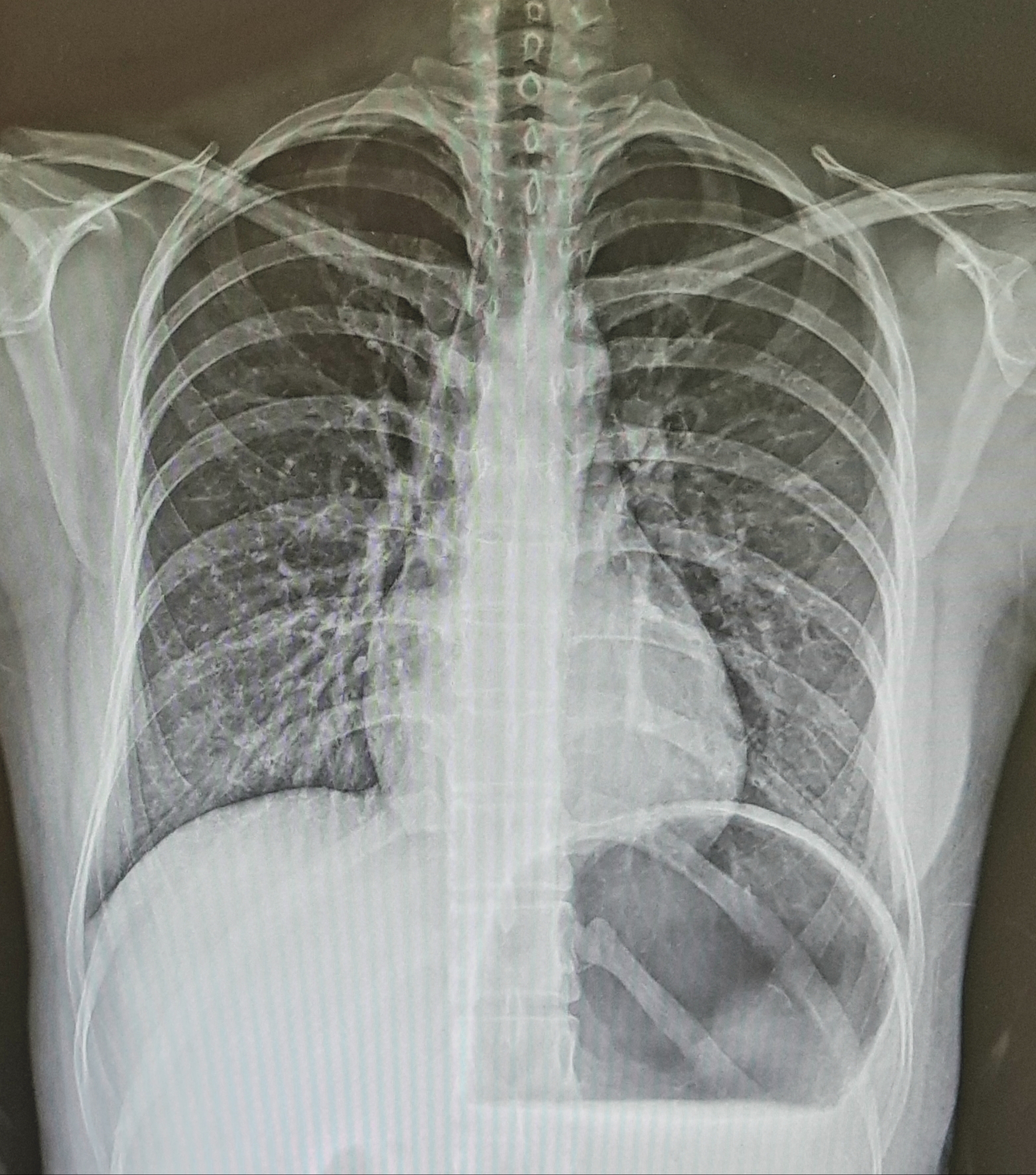
(영어/영문/English) 1. T1WI (T1 weighted image) To get a T1WI, a short TR and short TE are required. The clinically appropriate TR is 350~750ms, and the TE is 10~30ms. It is easy to see the anatomical structure and is mainly used in contrast imaging. 2. T2WI (T2 weighted image) To get a T2WI, long TR and long TE are needed. Clinically appropriate TR is over 2500ms and TE is 60~120ms. It is easy to i..
 [MRI] (영/한) Proton density weighted image - 양자밀도 강조영상
(영어/영문/English) The proton density weighted image is an image that obtains a contrast proportional to the number of protons in tissue existing within a patient. This image has the highest signal intensity and is mainly used to observe a wide range of tissues. When the proton density weighted image minimizes the T1 and T2 components as much as possible, a true proton density weighted image is obt..
 [MRI] (영/한) T2 Weighted Image (T2WI) - T2 강조영상
(영어/영문/English) Larmor frequency causes the longitudinal magnetization to lie on the horizontal plane (XY) , and then all spins in the same phase are rapidly dephased by interaction of spins and spins and inhomogeneity of the local magnetic field. (decay, incoherence, dephase, fan-out) It is the T2 weighted image that uses the phenomenon in which the spins in the transverse magnetization state a..
 [MRI] (영/한) T1 weighted image (T1WI) - T1 강조영상
(영어/영문/English) In the process of recovering the spins of the human body (recovering to the original state of the longitudinal magnetization) after receiving energy, the T1 weighted image is the image of the difference in T1 recovery time between tissues. The T1 weighted image is obtained by appropriately adjusting TR and TE. (the extrinsic factors representing the contrast of the image) The mos..