| 일 | 월 | 화 | 수 | 목 | 금 | 토 |
|---|---|---|---|---|---|---|
| 1 | 2 | 3 | 4 | 5 | ||
| 6 | 7 | 8 | 9 | 10 | 11 | 12 |
| 13 | 14 | 15 | 16 | 17 | 18 | 19 |
| 20 | 21 | 22 | 23 | 24 | 25 | 26 |
| 27 | 28 | 29 | 30 |
- slice gap
- TR TE
- aliasing artifact
- 사전포화펄스
- MR angiography
- FSE
- saturation band
- saturation pulse
- MRI 영상변수
- T2 이완
- T2WI
- fast spin echo
- 동위상 탈위상
- wrap around artifact
- 방사선사나라
- tof
- radiographer nara
- fractional echo
- T2강조영상
- T1WI
- MRA
- K-space
- MRI gantry
- ECG gating
- no phase wrap
- T1강조영상
- 자기공명혈관조영술
- receive bandwidth
- MRI image parameters
- chemical shift artifact
- Today
- Total
방사선사나라 Radiographer Nara
[MRI] (영/한) Image parameter(4) - Slice thickness , Slice gap / 영상변수(4) - 절편두께, 절편간격 본문
[MRI] (영/한) Image parameter(4) - Slice thickness , Slice gap / 영상변수(4) - 절편두께, 절편간격
SEONARA 2020. 4. 12. 14:37(영어/영문/English)
7. Slice thickness
Width of the slice to be scaned
Adjusting the width of the slice by passing a positive current to one side of the slice selection gradient coil and a negative current to the other side.
(If a high current is passed, the thickness of the slice becomes thin, and when a low current is passed, the thickness of the slice becomes thick.)
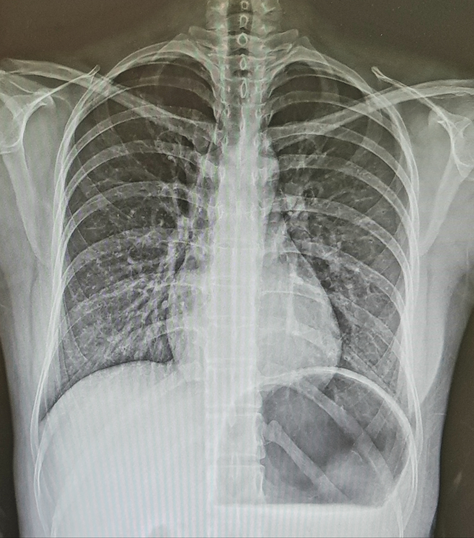
As the thickness of the slice increases, the SNR improves, but the partial volume effect of overlapping structures occurs and the spatial resolution decreases. Therefore, the slice thickness should be selected as thin as possible within a range where the anatomical structure of the tissue to be imaged does not overlap.
8. Slice gap
Gap between successive slices and slices
Ideally, it is desirable to minimize the slice gap, but if the image is obtained without giving the slice gap, the RF pulse excitations in an elliptical shape cause excessive interference with each other, resulting in image defects. As a result, the contrast and SNR of the image are lowered.
Therefore, in order to prevent such image defects, a certain slice gap should be provided, and an interval of about 25 to 50% of the slice thickness itself should be provided.
Alternatively, by converting an RF pulse close to an ellipse into an RF pulse close to a sinusoidal wave, the slice gap can be reduced by 10% without loss of image information and a change in scan time.

by radiographer nara
(국어/국문/Korean)
7. 절편두께
검사하고자 하는 단면의 폭
절편선택 경사코일의 한쪽에는 양의 전류를, 다른 한쪽에는 음의 전류를 흘려서 단면의 폭을 조절
(높은 전류를 흘리면 절편의 두께는 얇아지고 낮은 전류를 흘리면 절편의 두께는 두꺼워진다.)
절편의 두께가 증가할수록 SNR이 향상되지만 구조물들끼리 서로 겹치는 부분용적효과가 발생되고 공간분해능은 낮아진다. 그러므로 절편두께는 영상화하고자 하는 조직의 해부학적 구조가 겹치지 않는 범위에서 가능한 얇게 선택해야 한다.
8. 절편간격
연속적인 절편과 절편 사이의 공간
이상적으로는 절편 간격을 최소화하는 것이 바람직하지만 절편간격을 주지 않고 영상을 얻는다면 타원형 모양인 RF 펄스 여기가 서로 과도한 간섭을 유발하게 되고 이로 인해 영상결손이 발생한다. 이 결과로 영상의 대조도와 SNR이 낮아진다.
그러므로 이러한 영상결손을 예방하기 위해서는 어느 정도의 절편간격을 주어야 하는데 절편 자체두께의 25~50% 정도의 간격을 줘야 한다.
혹은 타원형에 가까운 RF 펄스를 정현파에 가까운 RF 펄스로 변환시켜 주면 영상정보의 손실과 검사시간의 변화 없이 절편간격을 10%까지 줄일 수 있다.
- 방사선사나라


