| 일 | 월 | 화 | 수 | 목 | 금 | 토 |
|---|---|---|---|---|---|---|
| 1 | 2 | 3 | ||||
| 4 | 5 | 6 | 7 | 8 | 9 | 10 |
| 11 | 12 | 13 | 14 | 15 | 16 | 17 |
| 18 | 19 | 20 | 21 | 22 | 23 | 24 |
| 25 | 26 | 27 | 28 | 29 | 30 | 31 |
- MRI gantry
- saturation band
- K-space
- T1강조영상
- FSE
- fractional echo
- 자기공명혈관조영술
- wrap around artifact
- TR TE
- T2강조영상
- 방사선사나라
- radiographer nara
- T2WI
- MRI 영상변수
- T2 이완
- MR angiography
- saturation pulse
- MRI image parameters
- no phase wrap
- aliasing artifact
- fast spin echo
- chemical shift artifact
- tof
- slice gap
- T1WI
- MRA
- 동위상 탈위상
- 사전포화펄스
- receive bandwidth
- ECG gating
- Today
- Total
방사선사나라 Radiographer Nara
[MRI](영/한) 3 Dimension Volume Image / 3-D 영상 획득 기법 본문
(영어/영문/English)
The concept of 3D in MR imaging refers to a method of acquiring images.
In the 3-dimension method, RF pulses are applied to the entire volume to be imaged to determine location information and measure the signal.
A 3D technique is possible by adding a slice encoding gradient in the direction of the slice selection gradient to the 2D method. The slice encoding gradient also changes with every TR, like the phase encoding gradient. The number of steps means the number of partitions.
For this reason, the scan time in 3D technique can be multiplied the number of slices by the scan time in 2D.
2D scan time = TR x Number of phase encoding x NEX
3D scan time = TR x Number of phase encoding x NEX x Number of slices
As such, the 3D technique takes a lot of scan time, so it is mainly used only in gradient echo with a short TR.
In the 3D technique, an image can be obtained without an gap, and the the number of signal measurements per partition increases, so that an image with good SNR can be obtained.
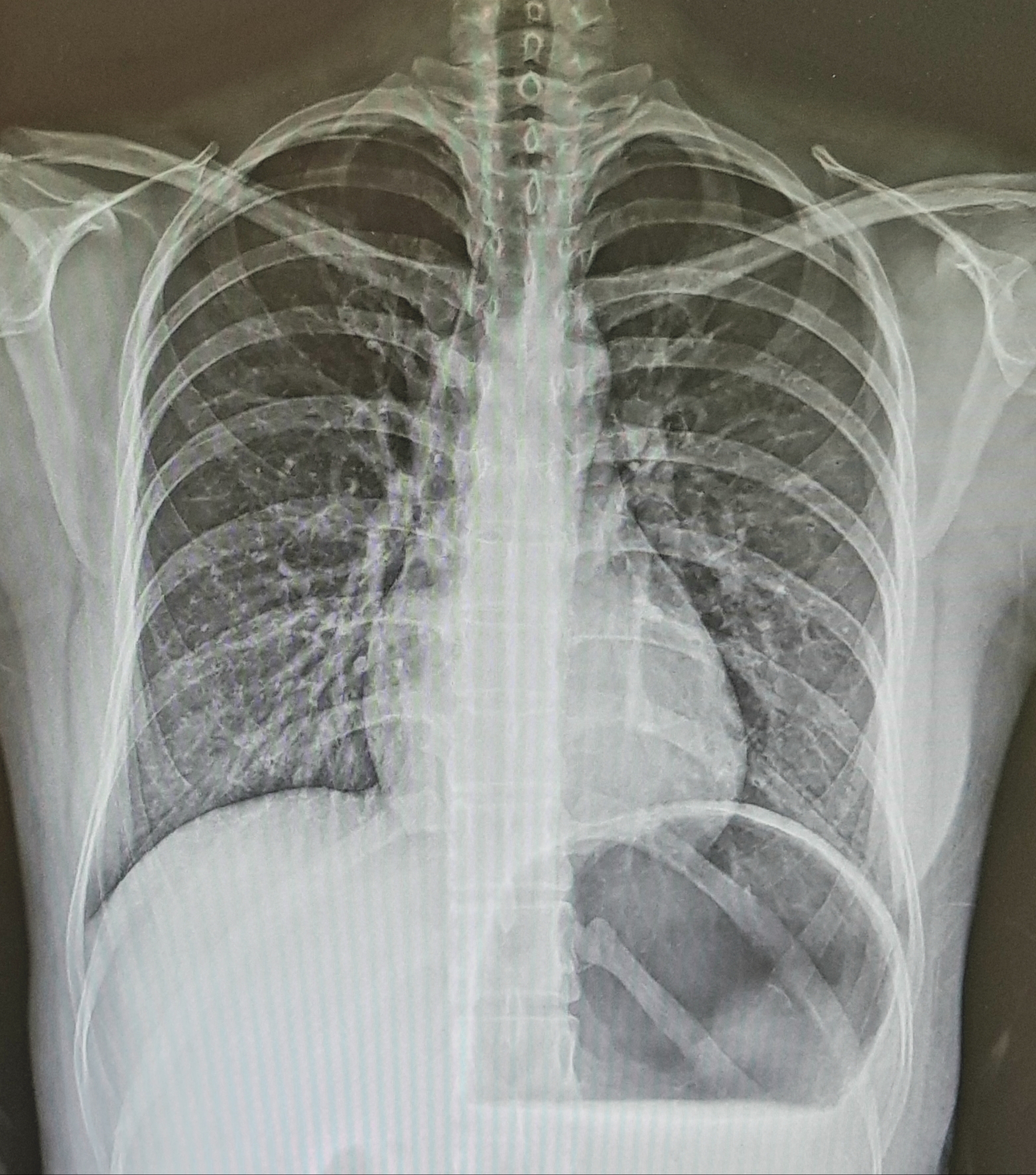
In clinical practice, it is mainly used for MR angiography, various joints, and cervical spine.

by radiographer nara
(국어/국문/Korean)
MR영상에서 말하는 3D의 개념은 영상을 획득하는 방법을 말한다.
3-dimension 방법은 영상화하려는 부피 전체에 RF 펄스를 인가하여 위치정보를 파악하고 신호를 측정한다.
2D 방법에 단면선택 경사자장 방향으로 경사자장을 추가함으로서 3D 기법이 가능하다. 단면 경사자장도 위상부호화 경사사장처럼 매 TR마다 변하는데 그 수가 단면의 수를 의미한다.
그렇기 때문에 3D기법에서의 검사시간은 2D에서의 검사시간에 단면수를 곱하면 된다.
2D 영상 검사시간 = TR x phase encoding 수 x NEX
3D 영상 검사시간 = TR x phase encoding 수 x NEX x 단면수
이와 같이 3D 기법은 검사시간이 많이 소요되므로 TR이 짦은 gradient echo에서만 주로 사용된다.
3D기법은 간격 없이 영상을 얻을 수 있고 각 단면당 신호 측정 수가 많아져 SNR이 좋은 영상을 얻을 수 있다.
임상에서는 주로 자기공명혈관조영술이나 각종 관절, 경추 등에 이용되고 있다.

- 방사선사나라
'자기공명영상 (MRI)' 카테고리의 다른 글
| [MRI](영/한) FSE (Fast Spin Echo), TSE(Turbo Spin Echo) / 고속 스핀에코 (0) | 2020.04.24 |
|---|---|
| [MRI](영/한) IR (Inversion Recovery) pulse sequence/ 반전회복 펄스 시퀀스 (0) | 2020.04.23 |
| [MRI](영/한) Gradient Echo의 종류 / 경사자계 에코의 종류 (0) | 2020.04.21 |
| [MRI] Gradient Echo (GRE) / 경사자계 에코 신호 (0) | 2020.04.20 |
| [MRI] (영/한) Spin Echo (SE) / 스핀에코 (0) | 2020.04.19 |


